


Technical Service Details of Multiplex Immunohistochemistry (mIHC)
Multiplex Immunohistochemistry (mIHC) technical service is a one-stop pathological detection service based on Tyramide Signal Amplification (TSA) technology. It enables simultaneous detection and quantitative analysis of multiple targets on a single tissue section, serving as a core tool for deciphering tissue microenvironments and disease mechanisms. Below is a detailed breakdown of mIHC technical service from seven dimensions: technical principle, core advantages, service content, sample requirements, service process, delivery standards, and application fields.
I. Technical Principle
mIHC achieves multi-target detection relying on Tyramide Signal Amplification (TSA) technology, with the core workflow as follows:
After the primary antibody binds to the target antigen, a secondary antibody conjugated with Horseradish Peroxidase (HRP) is added.
HRP catalyzes the activation of fluorophore-labeled tyramide molecules, which then covalently bind to tyrosine residues adjacent to the antigen, realizing signal amplification (the sensitivity is 10–1000 times higher than that of traditional IHC).
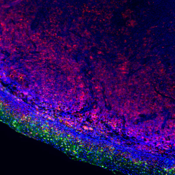
Unbound antibodies are eluted, and the above steps are repeated for multiple rounds of staining. Finally, simultaneous labeling of 2–7 targets (including DAPI) is accomplished on a single tissue section.
II. Core Advantages
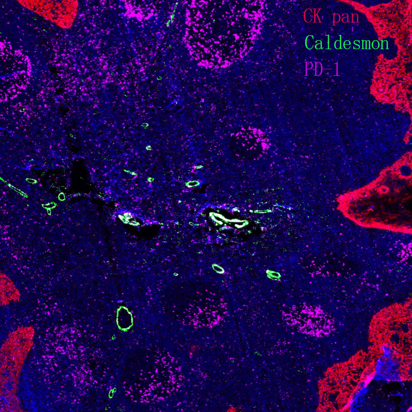
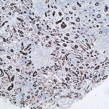
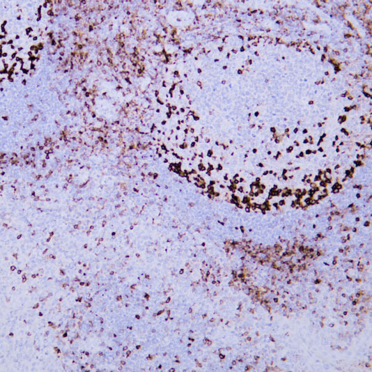
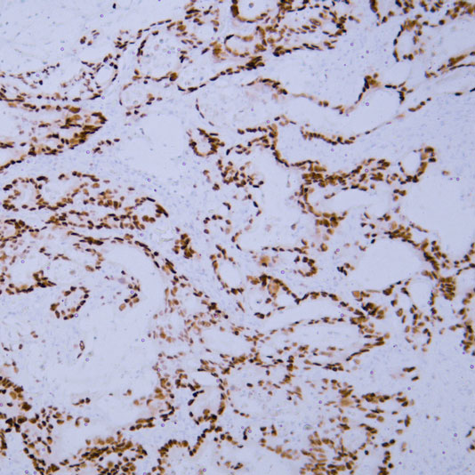
Multi-target in situ detectionA single section can achieve 2–7 color staining (maximum 6 targets with 7 colors), breaking the limitation of single-color detection in traditional IHC, and directly visualizing the spatial positional relationship among targets.
Ultra-high sensitivityThe signal amplification effect of TSA enables the detection of low-abundance targets, with sensitivity over 100 times higher than that of conventional methods.
No species restrictionPrimary antibodies derived from the same species can be used, avoiding species cross-reactivity and reducing the difficulty of antibody selection.
Quantitative analysis capabilityCombined with digital pathology scanning and image analysis software, it can output quantitative data such as target expression level, cell subset proportion, and colocalization coefficient.
Strong sample compatibilityIt is applicable to multiple sample types including paraffin-embedded sections, frozen sections, and Tissue Microarrays (TMA).
III. Service Content
mIHC technical service is divided into standardized packages and customized services. Mainstream service providers can offer 2–7 color detection schemes, with details as follows:
Service Name | Content Description | Experimental Cycle | Price |
Fluorescent Staining | Tissue single staining | 1 week | Inquiry |
Tissue double staining | 1 week | Inquiry | |
Tissue triple staining - TSA | 1–2 weeks | Inquiry | |
Tissue quadruple staining - TSA | 1–2 weeks | Inquiry | |
Tissue quintuple staining - TSA | 3–4 weeks | Inquiry | |
Tissue sextuple staining - TSA | 3–4 weeks | Inquiry | |
Tissue microarray fluorescent staining | 1–2 weeks | Inquiry | |
Imaging/Scaning | Confocal imaging | 1 week | Inquiry |
Tissue microarray scanning (white light) | 1 week | Inquiry | |
Regular section scanning (white light) | 1 week | Inquiry | |
Large tissue section scanning (white light) | 1 week | Inquiry | |
Fluorescent scanning of single-target two-color (regular size) | 1 week | Inquiry | |
Fluorescent scanning of double-target three-color (regular size) | 1 week | Inquiry | |
Fluorescent scanning of triple-target four-color (regular size) | 1 week | Inquiry | |
Fluorescent scanning of quadruple-target five-color (regular size) | 1 week | Inquiry | |
Fluorescent scanning of quintuple-target six-color (regular size) | 1 week | Inquiry | |
Fluorescent scanning of sextuple-target seven-color (regular size) | 1 week | Inquiry | |
Fluorescent scanning of tissue microarrays | 1 week | Inquiry |
In addition, other service requirements such as tissue microarray fabrication, digital section scanning, and image quantitative analysis (e.g., positive cell proportion, staining intensity, colocalization coefficient calculation) can be further negotiated.
IV. Sample Requirements
Clients are required to provide samples meeting the following standards. Specific requirements for different sample types are listed below:
Sample Type | Quantity Requirement | Preservation and Transportation |
Paraffin-embedded sections | (Number of test samples + 4) × Number of indicators | Room temperature preservation and transportation |
Paraffin blocks | Blocks containing intact tumor/target tissue | Room temperature preservation and transportation |
Frozen sections | OCT-embedded, 4–6 μm thick | -20℃ low-temperature transportation |
Antibody requirements: Clients need to provide primary antibodies by themselves (with antibody specification sheets and dilution ratios).
V. Service Process
mIHC technical service adopts a one-stop workflow with a standard cycle of 2–3 weeks. The specific steps are as follows:
Demand communication: Confirm target combinations, sample types, detection indicators, and analysis requirements.
Sample quality control: Test sample integrity and fixation quality; re-provision is required for unqualified samples.
Preliminary experiment: Optimize antibody dilution ratios and staining conditions to ensure signal specificity.
Formal experiment: Complete multi-color staining and section mounting according to the TSA protocol.
Scanning and analysis: Acquire high-resolution images using a digital pathology scanner and perform quantitative analysis with software.
Result delivery: Provide stained tissue sections, raw images, and analysis reports.
VI. Delivery Standards
The deliverables provided by service providers include physical samples and data files, with details as follows:
Physical samples: Stained tissue sections (paraffin-embedded/frozen).
Image data: Original images of 200× fields of view (3 regions, including lesion/paracancerous tissues), whole-slide scanned images, and single-color signal split images.
Analysis report: Including statistics of target expression levels, cell subset proportions, colocalization analysis results, and experimental methodology descriptions.
VII. Application Fields
mIHC technology is widely used in basic scientific research, drug development, and clinical diagnosis, with core application directions including:
Tumor microenvironment research: Analyze the spatial interaction between immune cells (T cells, macrophages) and tumor cells.
Immunotherapy mechanism research: Detect the co-expression of immune checkpoints such as PD-1/PD-L1 and CTLA-4.
Neuroscience: Neurotransmitter colocalization and glial cell subtype identification.
Drug development: Preclinical drug target validation and efficacy evaluation.
Clinical diagnosis: Tumor molecular typing and prognostic marker detection (e.g., tertiary lymphoid structure assessment in non-small cell lung cancer).
Related Resource Links


Related Promotional Journal Downloads


Explore Our Recommended Popular Products
More products

30,000+ high- quality products available online
Primary Antibodies, Secondary Antibodies, mIHC Kits, ELISA Kits, Proteins, Molecular Biology Products,Cell Lines,Reagents ...
Contact Us
-

400-801-6722
-

support@lamarck.cn
-


Order
-

Message