


Research on cell death mechanisms serves as the core hub for decoding the regulatory laws of life homeostasis, analyzing the pathological essence of major diseases, and constructing innovative therapeutic systems. Its significance spans three major fields: basic life sciences, disease mechanism research, and clinical translational medicine.
At the basic level, it reveals the underlying logic of embryonic development (e.g., interdigital cell apoptosis mediating digit differentiation), adult tissue homeostasis (e.g., clearance of billions of senescent/damaged cells daily), and innate immune defense (e.g., pyroptosis-mediated pathogen clearance), filling the cognitive gap in the complex regulatory networks of multicellular organisms.
At the disease level, abnormal cell death constitutes the common pathological basis for cancer (tumor cells evading apoptosis and adapting to resistance against novel cell death pathways), neurodegenerative diseases (excessive neuronal apoptosis/necroptosis), and inflammatory diseases (inflammatory dysregulation caused by pyroptosis/necroptosis), providing key targets for clarifying pathogenic mechanisms.
At the clinical level, it directly drives therapeutic innovation: it has not only spawned marketed or investigational drugs such as Bcl-2 inhibitors (for treating chronic lymphocytic leukemia) and GPX4 inhibitors (for inducing ferroptosis in tumors) but also enables circumvention of drug resistance in traditional therapies and enhancement of treatment specificity. In doing so, it provides fundamental theoretical support and technical pathways for addressing critical human health issues and unmet clinical needs.
Apoptosis is the most classic form of programmed cell death. For Western Blot (WB) detection, focus should be placed on the activation of the Caspase family, the balance of the Bcl-2 family, and mitochondrial pathway markers. The core is to distinguish between "full-length (inactive)" and "cleaved fragments (active)".
Marker Category | Specific Protein/Molecule | WB Detection Key Points |
Effector Caspases | Caspase-3, Caspase-7 | Detect cleaved fragments (Caspase-3: 17/19 kDa, full-length: 32 kDa). SDS-containing lysis buffer is recommended to avoid artificial activation. |
Initiator Caspases | Caspase-8, Caspase-9 | Caspase-8 (extrinsic pathway, cleaved fragments: 43/41 kDa); Caspase-9 (intrinsic pathway, cleaved fragment: 37 kDa). |
Bcl-2 Family | Bcl-2, Bax, Bak | Focus on the Bax/Bcl-2 ratio (pro-apoptotic/anti-apoptotic); non-reducing electrophoresis is required to detect Bak oligomerization. |
Mitochondrial Release Factor | Cytochrome C | Subcellular fractionation (separation of mitochondrial/cytoplasmic components) is necessary. Only the detection of signals in the cytoplasm indicates apoptosis activation. |
Late Apoptosis Marker | PARP-1 | Detect cleaved fragment (89 kDa, full-length: 116 kDa). Antibodies that recognize both full-length and cleaved forms are more accurate. |
Auxiliary Markers | Smac/DIABLO, XIAP | Cytoplasmic accumulation of Smac indicates mitochondrial damage; downregulation of XIAP (an anti-apoptotic protein) can assist in verifying apoptosis. |
The core of pyroptosis lies in the cleavage of the Gasdermin family and the release of inflammatory factors. WB detection should focus on "pore-forming proteins" and "inflammatory Caspase subtypes", while combining with the detection of inflammatory factors.
Marker Category | Specific Protein/Molecule | WB Detection Key Points |
Key Effector Proteins | GSDMD, GSDME | GSDMD (cleaved fragment: 31 kDa, full-length: 53 kDa); GSDME (cleaved fragment: 35 kDa, full-length: 50 kDa) — these are specific markers of pyroptosis. |
Inflammatory Caspases | Caspase-1, Caspase-4/5/11 | Caspase-1 (cleaved fragments: 20/10 kDa, full-length: 45 kDa); Caspase-4/5/11 (cleaved fragments: ~20 kDa). |
Inflammatory Factors | IL-1β, IL-18 | Detect mature forms (IL-1β: 17 kDa, pro-form: 31 kDa; IL-18: 18 kDa, pro-form: 24 kDa). Verification by ELISA is often required. |
Auxiliary Marker | NLRP3 | Detect protein expression level (full-length: 110 kDa). Note that its activation depends on oligomerization; WB can only reflect expression level, so co-immunoprecipitation is needed for assistance. |
Necroptosis is a "Caspase-independent" inflammatory cell death. The core of WB detection is the activation of the RIPK3-MLKL pathway, with a focus on detecting "phosphorylated modifications (active forms)".
Marker Category | Specific Protein/Molecule | WB Detection Key Points |
Core Kinases | RIPK1, RIPK3 | Detect phosphorylated forms (p-RIPK1: Ser166; p-RIPK3: Ser227/Thr231). The expression level of total protein has low reference value. |
Effector Protein | MLKL | The key is to detect phosphorylated MLKL (p-MLKL: Ser358). The active form translocates to the cell membrane, and subcellular localization can be used for assistance. |
Auxiliary Marker | Caspase-8 | Detect full-length (55 kDa). During necroptosis, Caspase-8 activity is inhibited, with no cleaved fragments (to avoid confusion with apoptosis). |
Ferroptosis is a form of cell death mediated by "iron-dependent lipid peroxidation". There are no "cleaved fragment" markers in WB detection; instead, focus on the expression of key regulatory proteins and "lipid peroxidation-related molecules".
Marker Category | Specific Protein/Molecule | WB Detection Key Points |
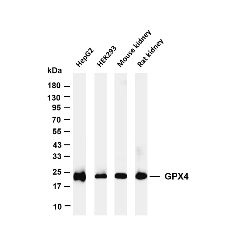
Core Regulatory Proteins | GPX4, SLC7A11 | GPX4 (22 kDa, downregulated expression is a core marker of ferroptosis); SLC7A11 (55 kDa, inhibition induces ferroptosis). |
Lipid Metabolism-Related | ACSL4, LPCAT3 | ACSL4 (72 kDa, promotes lipid peroxidation); LPCAT3 (50 kDa, maintains membrane lipid composition, sensitive to downregulation). |
Iron Metabolism-Related | FTH1, TFR1 | FTH1 (21 kDa, an iron storage protein; downregulation causes iron release); TFR1 (90 kDa, an iron uptake protein; upregulation indicates iron accumulation). |
Auxiliary Marker | Nrf2 | A key factor in the antioxidant pathway (65 kDa, activated and upregulated during ferroptosis). |
Cuproptosis is a form of cell death caused by "copper-dependent mitochondrial metabolic abnormalities". WB detection should focus on the FDX1-lipoylated protein pathway and "mitochondrial TCA cycle-related proteins".
Marker Category | Specific Protein/Molecule | WB Detection Key Points |
Core Regulatory Factors | FDX1, LIAS | FDX1 (20 kDa, a key mediator of copper toxicity; knockout rescues cuproptosis); LIAS (50 kDa, a lipoyl synthase; downregulation inhibits lipoylation). |
Lipoylation-Related Proteins | DLAT, PDHA1 | DLAT (51 kDa, a TCA cycle protein that aggregates after copper binding; soluble/insoluble fractions can be detected); PDHA1 (43 kDa, downregulated in lipoylation). |
Copper Transporters | SLC31A1, ATP7A/B | SLC31A1 (35 kDa, a copper importer; overexpression enhances copper sensitivity); ATP7A/B (160 kDa, copper exporters; downregulation causes copper accumulation). |
Auxiliary Marker | HSP70 | A heat shock protein (70 kDa, upregulated due to proteotoxic stress during cuproptosis). |
Autophagy is a "lysosomal degradation" process. The core of WB detection is LC3 lipidation, requiring the distinction between "LC3-I (free form)" and "LC3-II (membrane-bound form)".
Marker Category | Specific Protein/Molecule | WB Detection Key Points |
Core Marker | LC3B | Detect LC3-I (18 kDa) and LC3-II (16 kDa). An increased LC3-II/LC3-I ratio indicates autophagy activation; PE-conjugated antibodies are needed to enhance signals. |
Autophagy Regulatory Proteins | Beclin1, ULK1 | Beclin1 (60 kDa, a key factor in autophagy initiation; upregulation promotes autophagy); ULK1 (150 kDa, detect phosphorylated form p-ULK1: Ser555). |
Autophagy Substrate | p62/SQSTM1 | 48 kDa, degraded during autophagy activation (expression downregulated). Simultaneous upregulation with LC3-II indicates impaired autophagic flux. |
Lysosome-Related | LAMP1 | 110 kDa, a lysosomal membrane protein; upregulated expression indicates enhanced lysosomal activity, assisting in verifying autophagic flux. |
Related Resource Links


Related Promotional Journal Downloads


Explore Our Recommended Popular Products
More products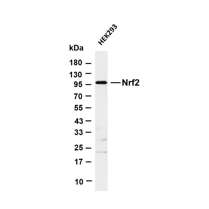

30,000+ high- quality products available online
Primary Antibodies, Secondary Antibodies, mIHC Kits, ELISA Kits, Proteins, Molecular Biology Products,Cell Lines,Reagents ...
Contact Us
-

400-801-6722
-

support@lamarck.cn
-


Order
-

Message