


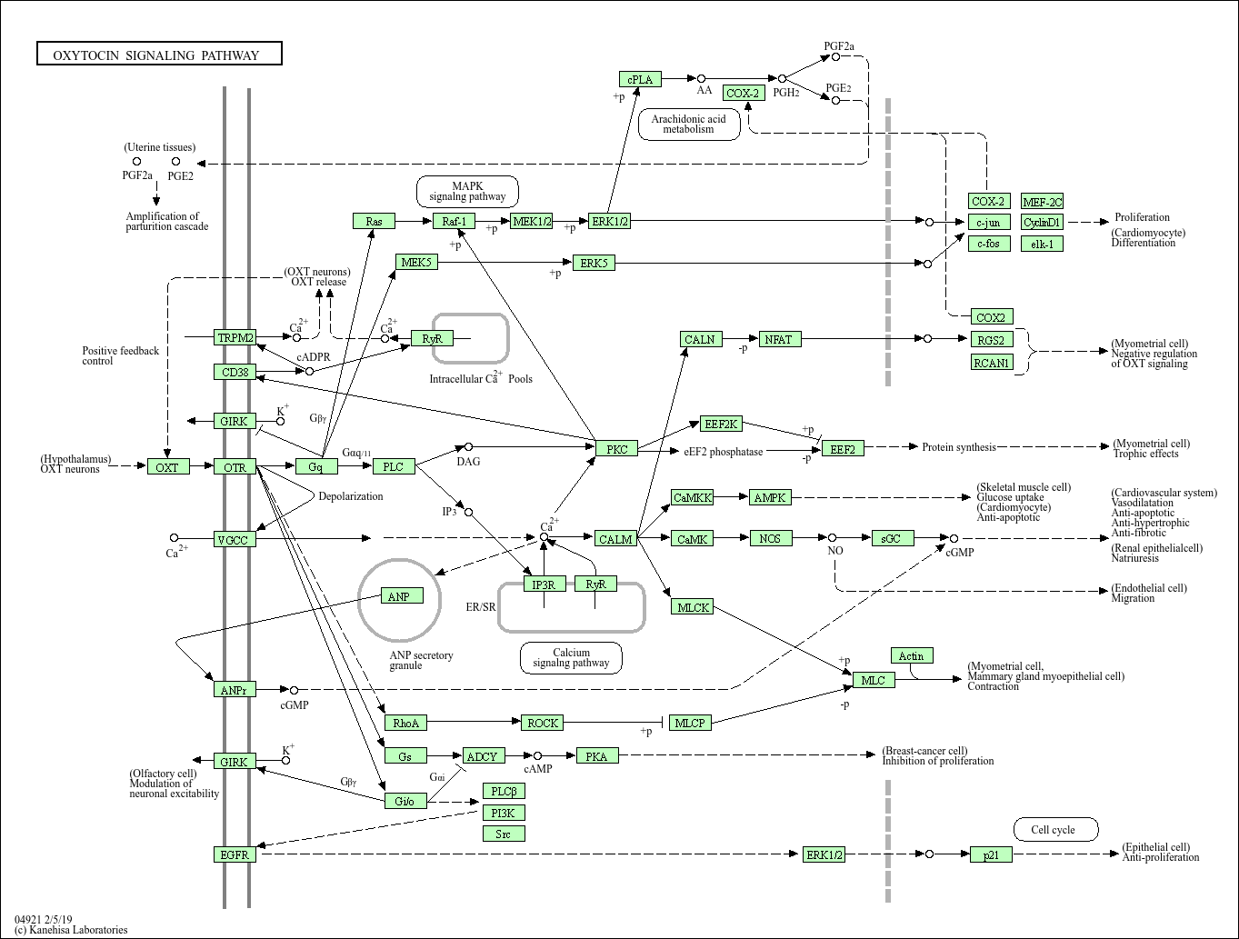
Oxytocin signaling pathway
Core of basic research: Focuses on the molecular mechanism by which oxytocin (OT) regulates uterine contraction, lactation reflex, and social behavior, a key pathway for reproductive physiology and social cognition. OT is synthesized by hypothalamic supraoptic and paraventricular nucleus neurons, stored in the posterior pituitary via the hypothalamic-pituitary tract, and released upon physiological stimuli (cervical dilation during childbirth, nipple stimulation during lactation). OT binds to Gq protein-coupled receptors (OTR) on target cells (uterine smooth muscle cells, mammary myoepithelial cells, central neurons), activating PLC-γ to hydrolyze PIP2 into IP3 and DAG. IP3 promotes endoplasmic reticulum calcium release, and DAG activates PKC, acting synergistically: in uterine smooth muscle cells, calcium binds calmodulin to activate myosin light chain kinase (MLCK), triggering myofilament sliding and uterine contraction; in mammary myoepithelial cells, calcium influx induces cell contraction to promote milk ejection. Additionally, OT regulates social behavior, anxiety, and parent-child attachment in the central nervous system. Research focuses on the neural regulation of OT release, tissue-specific expression of OTR, the association between calcium signaling and muscle contraction, and pathway abnormalities in labor disorders (uterine atony) and insufficient lactation.
Core key proteins: Oxytocin (OT), oxytocin receptor (OTR), Gq protein, PLC-γ (phospholipase C-γ), IP3R (endoplasmic reticulum calcium channel), PKC (protein kinase C), Ca²⁺ (calcium ion), CaM (calmodulin), MLCK (myosin light chain kinase), actin/myosin (smooth muscle contractile proteins), MAPK (ERK, involved in cell proliferation), PI3K/Akt (regulates cell survival), uterine smooth muscle cells, mammary myoepithelial cells, hypothalamic OT neurons.
Core key proteins: Oxytocin (OT), oxytocin receptor (OTR), Gq protein, PLC-γ (phospholipase C-γ), IP3R (endoplasmic reticulum calcium channel), PKC (protein kinase C), Ca²⁺ (calcium ion), CaM (calmodulin), MLCK (myosin light chain kinase), actin/myosin (smooth muscle contractile proteins), MAPK (ERK, involved in cell proliferation), PI3K/Akt (regulates cell survival), uterine smooth muscle cells, mammary myoepithelial cells, hypothalamic OT neurons.
Product list
-
{{item.title}}{{item.react}}{{item.applicat}}
Product list
Product name
Reactivity
Application
Related Resource Links


Related Promotional Journal Downloads


Explore Our Recommended Popular Products
More products

30,000+ high- quality products available online
Primary Antibodies, Secondary Antibodies, mIHC Kits, ELISA Kits, Proteins, Molecular Biology Products,Cell Lines,Reagents ...
Contact Us
-

400-801-6722
-

support@lamarck.cn
-


Order
-

Message